
Table of Contents
What is Gram Staining?
- Gram staining is a common technique used to differentiate two large groups of bacteria based on their different cell wall constituents.
- Gram staining is the most crucial staining in microbiology and is the first step in differentiating bacteria
- The Gram stain procedure distinguishes between Gram-positive and Gram-negative groups by coloring these cells into either red or violet.
- This method of Gram staining is named after, the Danish scientist Hans Christian Gram, who developed the technique while examining the lungs tissue from patient suffering from pneumonia
- Hans Christian discovered that certain stains were preferentially taken up and retained by the bacterial cell walls
- Counterstain was not used in this process which was later used by German pathologist Carl Weigert, added the final step in staining with Safranin
- Thus gram staining is the method of differentiating the bacterial species in two large groups (gram positive and gram negative)
Importance of Gram Staining
- Gram staining immediately helps to diagnose the presence or absence of bacterial infection.
- Gram staining is used to find out whether bacterial infection is present or not through identification of bacteria either gram positive or negative.
- Gram staining can also be used to identify fungal infections too.
- Gram staining helps the medical person/doctor to identify if a person has bacterial infection, and it determine what type of bacteria are causing it.
- Gram staining also helps to determine the effective treatment plan.
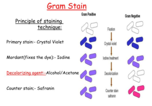
Reagents Used in Gram Staining
- Crystal violet as the primary stain
- Iodine solution/Gram’s iodine as the mordant
- A decolorizer made of acetone and alcohol (95%)
- Safranin as counterstain
Principles of Gram Staining
- The structure of the organism’s cell wall determines whether the bacteria is gram positive or gram negative
- When the bacteria is stained with primary stain and fixed by mordant, some bacteria are able to retain the primary stain by resisting the decolorization while other decolorized
- Those bacteria which retain the primary stain are Gram positive and those that don’t and get counterstain are Gram negative.
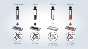
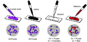
- The results of using different reagents are as follows:
1. Crystal violet: all bacteria takes crystal violet and all appear violet
2. Iodine: a complex is formed which is known as crystal violet iodine (CV-I)
3. Acetone/ethanol: bacteria with high lipid content loose CV-I complex and appear colorless but those with less lipid content retains CV-I complex and appear violet
4. Safranin: only the colorless bacteria take the color and appear pink
Procedure of Gram Staining
1. Preparation of slide smear
- A drop of suspended culture is transferred with the help of inoculation loop to the microscope slide.
- A drop of water is added to facilitate a minimal amount of colony transfer to the examination slide.
- A minimal amount of culture is required thus if culture can be detected visually on an inoculation loop, it shows there is too much culture.
- Culture is spread with an inoculation loop to an even thin film over a circle of 15mm in diameter.
- To dry the slide a gentle flame can be used by moving the slide circularly over the flame to prevent overheating or forming of ring patterns in the slide. It can also be air-dried
- The heat helps in proper cell adhesion to the glass slide and prevents the loss of culture during rinsing.
2. Gram staining
- Crystal Violet was poured and kept for about 30 seconds to 1 minutes
- After 10 to 60 seconds, the stain is poured off, and the excess stain is rinsed with water to wash off the stain without losing the fixed culture.
- Iodine solution is poured and covered the smear for 10 to 60 seconds which is known as “fixing the dye.” Iodine solution is poured off, and the slide is rinsed with running water. Excess water from the surface is taken off by shaking
- A few drops of decolorizer are added to the slide which is known as “solvent treatment.” And then slide is rinsed with water in 5 seconds. To prevent excess decolorization in the gram-positive cells, addition of decolorizer is stopped as soon as the solvent is not colored as it flows over the slide
- The smear is counterstained with safranin solution for 40 to 60 seconds. The safranin solution is washed off with water, and excess water is blotted with the bibulous paper. Air dry method can also be used after shaking off excess water
Summary of the Gram Staining Procedure
- Take a clean, grease free slide.
- Prepare the smear of suspension on the clean slide with a loopful of sample.
- Air dry and heat fix
- Pour the crystal violet and keep it for 30 seconds to 1 minute. Then, rinse with water.
- Flood the gram’s iodine for 1 minute and wash with water.
- Then ,wash with 95% alcohol or acetone for about 10-20 seconds and rinse with water.
- Add safranin for about 1 minute and wash with water.
- Air dry, blot dry and observe under Microscope.
Source https://www.researchgate.net/figure/Gram-staining-procedure-20_fig1_322681557
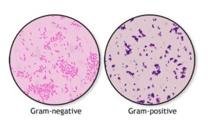
Interpretation
- Gram Positive bacteria: blue or purple color
- Gram Negative bacteria: red or pink color
Source: https://medlineplus.gov/ency/imagepages/19955.htm
Examples of Gram Positive and Gram Negative Bacteria
- Gram positive bacteria: Actinomyces, Bacillus, Clostridium, Corynebacterium, Enterococcus, Gardnerella, Lactobacillus, Listeria, Mycoplasma, Nocardia, Staphylococcus, Streptococcus, Streptomyces, etc.
- Gram negative bacteria: Escherichia coli (E. coli), Salmonella, Shigella, and other Enterobacteriaceae, Pseudomonas, Moraxella, Helicobacter, Stenotrophomonas, Bdellovibrio, acetic acid bacteria, Legionella etc.
Differences between Gram Positive and Gram Negative Bacteria
| Basis of differences | Gram Positive Bacteria | Gram Negative Bacteria |
| Gram reaction | Gram positive bacteria retain the crystal violet stain during gram staining | Gram negative bacteria do not retain the crystal violet stain during gram staining |
| Appearance under microscope | Gram positive bacteria appear purple or blue color after the gram staining | Gram negative bacteria appear red or pink in color after gram staining |
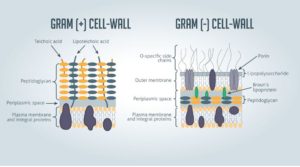
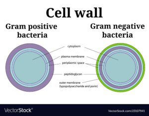
| Outer layer | Gram positive bacteria do not have outer cell wall beyond peptidoglycan membrane | Gram negative bacteria have hard, protective outer cell wall |
| Peptidoglycan layer | Peptidoglycan layer is thick, hard and multilayered | Peptidoglycan layer is thin and single layer |
| Periplasmic space | Periplasmic space is absent here | Periplasmic space is present here |
| Cell wall | Cell wall is about 20-80 nm with smooth structure containing virtually none lipopolysaccharide content | Cell wall is about 5-10 nm with wavy structure with high lipopolysaccharide content |
| Pili | Gram positive bacteria do not have pili | Gram negative bacteria consists of pili |
| Pathogenicity | Few of this type of bacteria are pathogenic | Most of this type bacteria are pathogenic |
| Antibiotic resistance | They are more susceptible to antibiotic resistance such as penicillin and sulfonamide | They are more resistant towards antibiotics |
References and For More Information
https://www.ncbi.nlm.nih.gov/books/NBK562156/
https://asm.org/Protocols/Gram-Stain-Protocols
https://www.webmd.com/a-to-z-guides/difference-between-gram-positive-bacillus-gram-negative-bacillus
https://www.technologynetworks.com/immunology/articles/gram-positive-vs-gram-negative-323007
https://asm.org/Protocols/Gram-Stain-Protocols